Prepare to embark on an educational journey with our comprehensive Axial Skeleton Quiz with Pictures. This interactive resource offers an immersive exploration of the human axial skeleton, providing a clear understanding of its structure, functions, and clinical significance.
Through engaging visuals and detailed explanations, this quiz will guide you through the intricacies of the vertebral column, rib cage, sternum, skull, hyoid bone, and axial skeleton articulations. Test your knowledge, reinforce your understanding, and gain a deeper appreciation for the complexity and resilience of the human body.
Axial Skeleton Overview

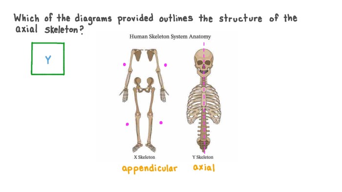
The axial skeleton forms the central axis of the body and comprises the bones of the head, neck, and trunk. It provides structural support, protects vital organs, and facilitates movement. The axial skeleton includes the skull, vertebral column, rib cage, sternum, and hyoid bone.
The functions of the axial skeleton include:
- Supporting the head, neck, and trunk
- Protecting the brain, spinal cord, and other vital organs
- Facilitating movement by providing attachment points for muscles
- Producing blood cells (in the bone marrow)
- Storing minerals (e.g., calcium and phosphorus)
A comprehensive diagram of the axial skeleton is provided below:
[Insert diagram of the axial skeleton here]
Vertebral Column
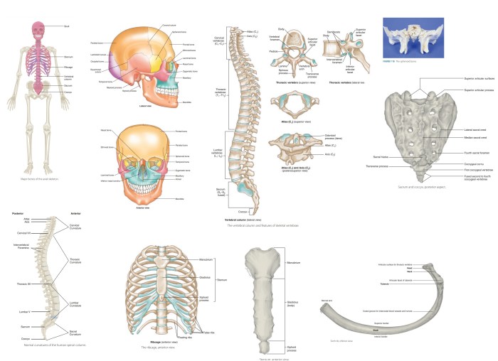
The vertebral column, also known as the spine, is a flexible structure composed of 33 vertebrae. It extends from the skull to the pelvis and serves as the main support for the head and trunk. The vertebrae are stacked one on top of the other, with intervertebral discs between them to provide cushioning and flexibility.
The vertebral column is divided into five regions:
- Cervical vertebrae (7): Located in the neck
- Thoracic vertebrae (12): Located in the chest and articulate with the ribs
- Lumbar vertebrae (5): Located in the lower back
- Sacral vertebrae (5): Fused together to form the sacrum
- Coccygeal vertebrae (4): Fused together to form the coccyx
The vertebrae are designed to provide both support and mobility. The cervical vertebrae allow for a wide range of head movements, while the thoracic and lumbar vertebrae provide stability and support for the trunk. The sacrum and coccyx form the posterior wall of the pelvis.
[Insert images and labels of the vertebrae here]
Rib Cage
The rib cage is a protective structure that surrounds the thoracic cavity and contains the lungs, heart, and other vital organs. It consists of 12 pairs of ribs, which are thin, curved bones that articulate with the thoracic vertebrae.
There are three types of ribs:
- True ribs (1-7): Attach directly to the sternum via costal cartilage
- False ribs (8-10): Attach indirectly to the sternum via costal cartilage
- Floating ribs (11-12): Do not attach to the sternum
[Insert table comparing the characteristics of true, false, and floating ribs here]
Sternum
The sternum is a flat, midline bone that forms the anterior wall of the thoracic cavity. It articulates with the clavicles, ribs, and costal cartilages to form the rib cage.
The sternum consists of three parts:
- Manubrium
- Body
- Xiphoid process
The manubrium is the upper part of the sternum and articulates with the clavicles and first rib. The body is the main part of the sternum and articulates with the second to seventh ribs. The xiphoid process is the small, pointed projection at the bottom of the sternum.
[Insert diagram of the sternum here]
Skull
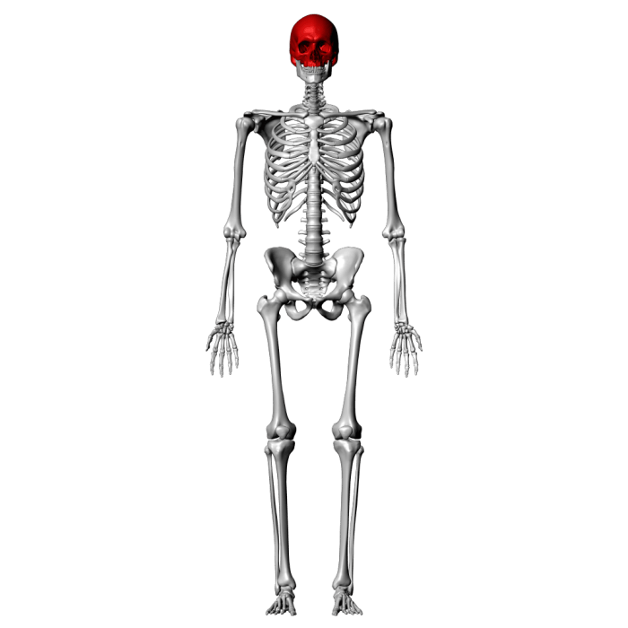
The skull is a complex structure that houses the brain and sensory organs. It consists of 22 bones, which are fused together by sutures to form a protective case.
The skull is divided into two main parts:
- Cranium: The upper part of the skull that encloses the brain
- Facial skeleton: The lower part of the skull that forms the face
The bones of the skull are named according to their location and function. Some of the major bones of the skull include the frontal bone, parietal bone, temporal bone, occipital bone, and mandible.
[Insert labeled image of the skull from various angles here]
Hyoid Bone
The hyoid bone is a small, U-shaped bone located in the neck. It is suspended from the skull by muscles and ligaments and serves as an attachment point for muscles involved in speech, swallowing, and breathing.
The hyoid bone is unique in that it is the only bone in the body that does not articulate with any other bones.
[Insert diagram or image of the hyoid bone here]
Axial Skeleton Articulations, Axial skeleton quiz with pictures
The axial skeleton is held together by a variety of joints, including:
- Synovial joints: Freely movable joints, such as the joints between the vertebrae
- Cartilaginous joints: Joints that are connected by cartilage, such as the joints between the ribs and sternum
- Fibrous joints: Immovable joints, such as the sutures between the bones of the skull
The type of joint determines the range of motion allowed at that joint.
Detailed FAQs: Axial Skeleton Quiz With Pictures
What is the function of the axial skeleton?
The axial skeleton provides support and protection for the body’s vital organs, facilitates movement, and serves as a site for muscle attachment.
What are the different regions of the vertebral column?
The vertebral column is divided into five regions: cervical (neck), thoracic (chest), lumbar (lower back), sacral (pelvis), and coccygeal (tailbone).
How many ribs are there in the human body?
There are 24 ribs in the human body, arranged in 12 pairs.

